The study and interpretation of visual fields is a very important part of ophthalmology and used for many different conditions, ranging from glaucoma (its most common use) to localizing neurological defects, and even to evaluate some retinal diseases. There are many different methods for assessing visual fields that the ophthalmologist has to be familiar with - not only do we have to be proficient at performing some of these tests ourselves, but we need to know appropriate indications and what clinical situations might warrant one technique above another.
In this series, we'll discuss the important aspects of visual fields, the techniques, and practical (and potentially testable) points.
Before we get started with the individual techniques of measuring visual fields, it's important to understand some basic terms and principles. Entire books have been written about visual fields, so while this and similar articles are geared towards basic review, you may wish to check out some of the resources below to get more details.
Mapping The Hill
If you read pretty much any text about visual fields, there's this classic description of visual fields as an "island or hill of vision in a sea of darkness," coined by Traquair in 1927 (1). You may have even seen figures showing the "hill" (2).
Here is a webpage discussing visual fields and demonstrating the "island of vision":
If you're anything like me, it's taken me multiple readings and countless re-readings to form the thousand words this picture depicts so neatly. This article is my meager attempt to synthesize how I learned the significance of this oft-depicted diagram, and to flesh out some terminology that is tossed out frequently in the field of perimetry (the measurement of visual field function).
What is the "hill of vision?"
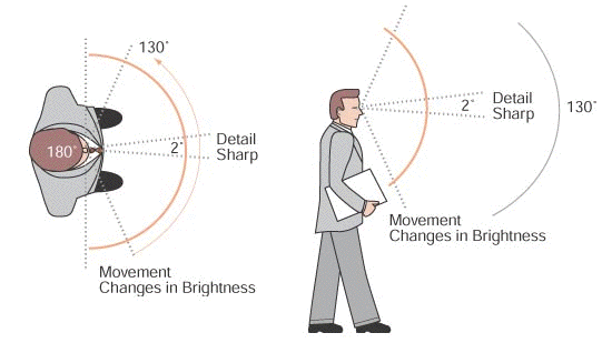
The "hill" or "island" of vision is a visual description of how our vision and visual fields work. We don't have high-definition vision in all areas of our visual field - in fact, there's only a very narrow portion of our visual field that provides high-resolution images. This is represented by the apex of the hill/island. Another way to consider it is how sensitive our vision is.
Visual sensitivity describes the level of detail that you are able to perceive; in other words, how small you can make a point and still be able to see it. As you might expect, the highest visual sensitivity is at fixation, which corresponds to the foveola, and is depicted on the diagram as the sharp peak. Because the foveola is such a small portion of the overall retina, the region of highest visual sensitivity is very narrow. As you move further away from the foveola, the visual sensitivity decreases (i.e., you need a larger target in order to see it) but the visual field extent is much larger (see below). Visual sensitivity is typically determined by using various techniques to find the smallest or dimmest (or both) object that can be seen at any given point in the visual field. That minimum level of vision is referred to as the threshold.
In general, there is a fairly steep drop-off in how detectable a test object is from fixation, with a gradual sloping of similar levels of visual sensitivity towards the periphery. The borders of the hill/island demarcate the extent of the visual field.
The extent of the visual field refers to how far you can move a target away from fixation and still be able to see it. For example, large and bright targets can be seen further in the periphery of our vision than small and dim targets. When this is plotted out in 3 dimensions, the diagram you see above forms. If mapped out in 2 dimensions, the vertical/z-axis in the 3-D diagram, which refers to the visual sensitivity, is usually displayed as concentric lines encircling fixation. Each of these lines is called an isopter.
Here are some conceptual take-aways:
- A small and dim object can be within our theoretical extent of vision, but be completely undetectable to us. I tried to use this fact to explain why I couldn't see all of the dirty dishes lying around the house, but my wife wouldn't buy it.
- The larger and brighter the object, the more likely it is for us to see it anywhere in our peripheral vision. This is why shiny objects grab our attention...probably.
- The area of our clearest, sharpest, and most sensitive vision is actually quite narrow. Even a small macular lesion can cause a very noticeable drop in acuity.
Normal Visual Field Extent
Visual fields are often described to be within x degrees of fixation or expressed as a diameter such as “central 30°,” which would correspond to a circle with a 30° radius from fixation. It’s helpful to know the generally accepted “normal” visual field extent (not factoring visual sensitivity), which corresponds to the retinal anatomy.
Visual Field Quadrant
Superior
Nasal
Inferior
Temporal
Degrees From Fixation (3)
50°
60°
70°
100°
If you sum these numbers, you theoretically should have about a total of 120° of vertical visual field extent, and around 160° of horizontal extent.
Diagram of horizontal and vertical visual field extents.
Image from the University of Edinburgh, Scottish Sensory Centre.
There are several ways that I’ve used to remember these facts. As you might expect, the superior and nasal visual fields are more narrow than the inferior and temporal visual fields (due to the eyelids and nose); thus, one way to remember the order might be the acronym SNIT (superior < nasal < inferior < temporal). Of course, this does not help you remember the actual numbers; hopefully the numbers aren't too tricky to memorize on their own. And if you have any suggestions on helping to remember the numbers, email me or leave a comment!
If there's any take-home point about these numbers, it's that most of the techniques that we use to assess visual fields don't actually test the far reaches of our visual field extent. In fact, even a Humphrey 30-2 visual field "only" tests the central 30° (30° on each side of fixation!) - meaning that further peripheral visual field defects theoretically can go undetected unless they eventually progress to involve fixation. However, because there is NOT 1-for-1 mapping of the visual fields to the occipital cortex, a central 30° test will map most of our functional visual field (in other words, most of the occipital cortex is used for central vision, so missing the far peripheral vision isn't a huge deal). For practical purposes, only kinetic perimetry (of which Goldmann perimetry is an example) tests the entire visual field.
Additional Reading and References
- Walsh TJ, ed. 2011. Visual Fields: Examination and Interpretation. 3rd Edition. Oxford: American Academy of Ophthalmology.
- Liu GT, Volpe NJ, Galetta SL, eds. 2010. Neuro-Ophthalmology: Diagnosis and Management. 2nd Edition. China: Elsevier.
- Miller NR, Newman NJ, Biousse V, Kerrison JB, eds. 2005. Walsh and Hoyt's Clinical Neuro-Ophthalmology. 6th Edition. Philadelphia: Lippincott Williams & Wilkins.
- [AAO] American Academy of Ophthalmology. 2015. Basic and Clinical Science Course, Section 5: Neuro-Ophthalmology. San Francisco: AAO.
- [AAO] American Academy of Ophthalmology. 2015. Basic and Clinical Science Course, Section 10: Glaucoma. San Francisco: AAO.
- Piltz-Seymour JR, Heath-Phillip O, Drance SM. Visual Fields in Glaucoma. Volume 3, Chapter 49. In: Tasman W, Jaeger EA, eds. 2006. Duane's Ophthalmology on CD-ROM. 2006 Edition. Online.
- Traquair HM. An Introduction to Clinical Perimetry. 5th ed. London, Henry Kimpton, 1946. pp 1-16.
- Piltz-Seymour JR, Heath-Phillip O, Drance SM. Visual Fields in Glaucoma. Volume 3, Chapter 49. In: Tasman W, Jaeger EA, eds. 2006. Duane's Ophthalmology on CD-ROM. 2006 Edition. Online.
- [AAO] American Academy of Ophthalmology. 2015. Basic and Clinical Science Course, Section 10: Glaucoma. San Francisco: AAO.
Do you have any specific questions about visual fields? How did you learn the basic concepts of visual fields? Leave a comment or send us an e-mail (ophthreview [at] gmail [dot] com)!


