Warning: This article, while intended to be read by ophthalmologists of all stages of training and practice, is extremely detailed. It started out as a small literature review on a topic I see in clinic quite a bit, and devolved into a really long post. While it may be less "review"-like than some of the other test-review articles I write, hopefully this clarifies a topic that was quite confusing to me when I was going through residency.
I have to be honest, I have been nervous venturing into the realm of the detailed literature review on this site. As I've stated before, I am still pretty fresh out of training and while I aspire to grow this site as part of my own collection of thoughts and notes on learning the entirety of ophthalmology, I don't always feel qualified to speak with authority on various topics. Additionally, as a fellowship-trained neuro-ophthalmologist, I recognize that I will be more likely to find interest in writing more detailed articles about neuro-ophthalmology, sometimes at the expense of other topics. Part of that is my own self-interest, but the other part is feeling qualified to know or develop sufficiently detailed literature review in other subjects. For example, while I want to keep up my knowledge of corneal disease, I know that my depth of knowledge of corneal disease will likely never be as much as a fellowship-trained cornea surgeon. Hopefully in the future, as I continue to build the site around the very basic topics, I will be able to recruit some contributors in different subspecialties to help provide the depth of understanding that I lack in those areas. I'm also very open to input from you readers, since I want to make sure that while I am primarily building a website for my personal study, I can also provide useful material for you all as well.
Introduction

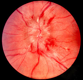
Papilledema. No one wants to see this on a Friday afternoon.
Image credit: Wikipedia
Pseudotumor cerebri syndrome (PTC, also referred to as idiopathic intracranial hypertension [IIH]) is classically taught as presenting in young, overweight women of childbearing age, with a history of headaches and findings of bilateral optic nerve swelling, associated with an elevated intracranial pressure. However, as with every "textbook" definition of a disease, there are atypical cases (children, men, thin people, older people), and so I am often confronted with some interesting diagnostic challenges when I am referred a patient that does not fit the typical picture of PTC who has bilateral optic nerve swelling.
Of course, not every overweight young female with a headache has PTC. And not all optic nerve swelling is papilledema. And while I think that any patient with either should have a detailed funduscopic examination and at least have PTC in the differential diagnosis, some basic principles can be applied to help determine if those patients need further evaluation. Also, for those of us who are either studying for ophthalmology exams or don't have the luxury of having a neuro-ophthalmologist nearby to help, it's useful to know what to do with these patients.
There are a lot of different aspects to cover with PTC, which I plan to eventually discuss (similar to my plans to cover the entire breadth of ophthalmology, even if it takes me the next 20 years). So stay tuned for more articles covering PTC in the future!
As we learn more and more about PTC, a greater emphasis has been made within the neuro-ophthalmology community to tighten up the diagnostic criteria for PTC, which in turn helps us determine the appropriate treatment options.
Naming Confusion?
Since Quincke first described the condition in 1897 (1), there have been many names for this condition, including toxic hydrocephalus (2), otitic hydrocephalus (3), hypertensive meningeal hydrops (4), pseudoabscess (5), ICP without brain tumor (6), brain swelling of unknown cause (7-8), and papilledema of indeterminate etiology (9). In 1914, Warrington coined the term "pseudotumor cerebri," which remains the most popular name for this condition (10).
Foley introduced "benign intracranial hypertension" in 1955 (11) (it remains the default terminology in ICD-9 and ICD-10) (12), but this name is now strongly discouraged, since "benign" implies no permanent or devastating sequelae, and in upwards of 25% of patients with IIH will have severe debilitating visual loss (13-15). In fact, several well-respected texts (including Walsh and Hoyt's Clinical Neuro-Ophthalmology by Miller et al and Neuro-Ophthalmology: Diagnosis and Management by Liu et al) acknowledge the historical role of this term, but emphasize its shortcomings (16-17). Bottom line, don't call it benign intracranial hypertension!
In more recent times, pseudotumor cerebri and idiopathic intracranial hypertension have become more widespread terms for similar (but not always synonymous) conditions. Dr. Deborah Friedman, one of the many well-published experts on the subject, makes the distinction between the two terms in this respect:
[Dr. Friedman and her colleagues] propose that patients can be subdivided into those with primary vs secondary PTC. IIH is a subset within the primary PTC category, while the secondary PTC group would include causes such as venous sinus thrombosis, medications, and medical conditions (18).
In other words, pseudotumor cerebri is the "umbrella" term used to describe the syndrome of increased intracranial pressure regardless of etiology, and idiopathic intracranial hypertension is the term used to describe increased intracranial pressure without any known cause.
Why Does This Even Matter?
I'm sure many of you who have clicked through this article are wondering, why the long history lesson? After all, if most people use PTC and IIH interchangeably, does it even matter what we call it?
Well, to understand the rationale behind the current diagnostic criteria for PTC, we need to at least consider the evolution of terminology. Just as papilledema no longer means optic nerve swelling regardless of etiology, we need to have a common language of terms to avoid any major confusion.
Thus, for neuro-ophthalmologists (and should be for everyone else too), it is incorrect to describe a patient with increased intracranial pressure from a cerebral venous thrombosis or minocycline use as having "IIH" (since there is a secondary cause it cannot be idiopathic), but it is perfectly valid to diagnose them as having "pseudotumor cerebri secondary to _____" or "intracranial hypertension secondary to _____." While it seems a bit nitpicky, it's important to recognize the difference here, since it allows us to properly interpret and apply the clinical research (since some studies look solely at patients with IIH, and others at the broader PTC syndrome).
Diagnostic Criteria For Pseudotumor Cerebri: A History
The Modified Dandy Criteria
In 1985, Dr. J. Lawton Smith proposed the modified Dandy criteria for diagnosis of PTC. These include (19-20):
- Presence of symptoms and signs of increased intracranial pressure
- No localizing neurologic signs
- Cerebrospinal fluid studies demonstrating elevated opening pressure and normal CSF studies
- Small or normal cerebral ventricles noted on neuroimaging
The Modified-Modified Dandy Criteria
In recent years, there have been several major articles discussing the diagnostic criteria for IIH. Friedman and Jacobson provided the "modified-modified Dandy criteria" in 2002, which expanded the language of the previous criteria detailed by Smith (16-17, 21):
- Symptoms or signs must be solely attributed to intracranial hypertension or papilledema.
- Documented elevated intracranial pressure (ICP) measured in the lateral decubitus position.
- Normal CSF composition.
- Normal neuroimaging (found on MRI or contrast-enhanced CT for typical patients, and including MRV for atypical patients).
- No other cause of increased ICP found.
The 2013 Revised Diagnostic Criteria: Friedman, Liu, Digre (18)
In 2013, Friedman, Liu, and Digre revisited the diagnostic criteria to include new information about neuroimaging, reports of pseudotumor cerebri without papilledema, and many other clinical similar conditions that may be lumped into the new PTC syndrome umbrella.
The authors noted that those with true primary pseudotumor cerebri syndrome (i.e., IIH) tended to be older than 3 years old and younger than 60 years old. So although the patient's age is not listed as a strict requirement for diagnosis, alternate causes for optic nerve swelling or increased intracranial pressure should be considered in those extreme outliers.
They divided the criteria into "PTC with papilledema" and "PTC without papilledema." Additionally, because patients with PTC can have normal intracranial pressures at times (22), a patient with a high degree of suspicion for PTC but with a normal intracranial pressure on lumbar puncture may still be diagnosed with "probable PTC" and managed based on clinical symptoms. However, all of the other criteria still need to be satisfied (adapted from Friedman et al):
- Papilledema must be present
- Normal neurological examination except for cranial nerve abnormalities
- Neuroimaging: normal brain parenchyma without evidence of hydrocephalus, mass, or structural lesion and no abnormal meningeal enhancement on MRI, with and without gadolinium, for typical patients (female and obese), and MRI, with and without gadolinium, and magnetic resonance venography for others; if MRI is unavailable or contraindicated, contrast-enhanced CT may be used
- Normal CSF composition
- Elevated lumbar puncture opening pressure (≥250 mm CSF in adults and ≥280 mm CSF in children [250 mm CSF if the child is not sedated and not obese]) in a properly performed lumbar puncture
If papilledema is not present, then there must either be a CN6 palsy (unilateral or bilateral) or ≥ 3 neuroimaging criteria satisfied:
- Empty sella
- Flattening of the posterior aspect of the globe
- Distention of the perioptic subarachnoid space with or without a tortuous optic nerve
- Transverse venous sinus stenosis
There are several key takeaway points from this article:
- Headaches, transient visual obscurations, pulsatile tinnitus, blurred vision, and any other symptoms previously described in papilledema or PTC/IIH are no longer valid criteria for the diagnosis of PTC/IIH. Additionally, while PTC/IIH is classically described in the obese young female, it is not a requisite for diagnosis.
- Absence of spontaneous venous pulsations, while somewhat helpful in description of the intracranial pressure, is not sufficiently diagnostic. While the presence of spontaneous venous pulsations is highly suggestive of intracranial pressure < 190 mmCSF, it is not definitive (23).
- Cranial neuropathies other than CN6 palsy are rare in intracranial hypertension, but have been reported (24).
- The opening pressure has to be pretty high in order to have a definitive diagnosis of PTC/IIH. I had a pediatric patient who presented to me diagnosed with a "high" opening pressure of 180 mmCSF once - this criteria helps me know when to diagnose someone with definite PTC/IIH, probable PTC/IIH, and to rule it out.
- Both neuroimaging and lumbar puncture opening pressure are required for diagnosis of PTC/IIH.
Putting It Together: How To Diagnose Pseudotumor Cerebri
So now that we have a (hopefully) comprehensive look at the diagnostic criteria for PTC, how do we translate that information into our clinical situation? Let's walk through a few case examples. Note that I am only going to discuss the process to diagnosis, not treatment (that will likely be at least 1 other article): (Caveat: these are generalized principles and not prescriptive for every situation. This is not medical advice!)
Case 1:
23-year old obese female with no significant past medical history, a 4-month history of headaches, and no visual complaints presents for a routine vision exam. She reports a 20-kg weight gain over the past 6 months. Upon further questioning, she complains of some transient visual obscurations and pulsatile tinnituse but no diplopia. Best-corrected visual acuity is 20/20 in each eye, with normal visual fields to confrontation and no relative afferent pupillary defect. Her motility and alignment are normal. Optic nerves show bilateral swelling suggestive of papilledema. How do you evaluate this patient?
In many ways, this is your textbook case of PTC. This is a young obese female with headaches and classic symptoms of papilledema and increased intracranial pressure. Generally the optic nerve swelling is bilateral and in the setting of normal vision and visual fields. There are no other cranial neuropathies reported.
So what IS the next step? In many cases, I have seen patients sent directly to the ER for testing. Within the neuro-ophthalmologic community in the U.S., these cases are generally managed as an outpatient, with the rare exception being sent to the ER (perhaps they have significant visual field loss or some other symptom or exam finding that would suggest a more fulminant course). Of course, the standard of care for evaluation of papilledema may vary depending on location. Here is one way to consider evaluating the patient, based on the information provided to us by the articles summarized above (and with some help from Neuro-Ophthalmology: Diagnosis and Management) (17):
Initial visit (you are the first to see the papilledema, patient already dilated):
- MRI brain with and without gadolinium (if not possible, then CT brain with contrast). I am going to refrain from spelling out a specific time frame for how soon this needs to be done in relation to the initial visit for several reasons: 1) the urgency of testing is largely framed within the context of active vision loss or threat of vision loss (in this case, she has a lower risk), and 2) depending on the resources available to you, it may not be practical to order an MRI or CT urgently. The MRI should be performed prior to the lumbar puncture. Additionally, an MRI is the preferred test over a contrast-enhanced CT. A non-contrast CT does not meet the criteria listed above for adequate neuroimaging.
- Lumbar puncture, performed in the lateral decubitus position, with opening pressure. Some patients, for various reasons, may get lumbar punctures in non-ideal positions. Unfortunately, when this happens it is much more difficult to interpret the opening pressure. There is no direct mathematical conversion between opening pressures measured in the lateral decubitus position vs. those measured in the prone position vs. those measured in the sitting position. As such, the order (assuming that you are not performing the LP yourself) needs to specify that you want the procedure done in the lateral decubitus position. Here are a few other helpful hints on how to order the lumbar puncture:
- Generally, neurologists and interventional radiologists routinely perform lumbar punctures. Depending on the resources available to you, it may be helpful to develop a good referral relationship between one or both of these specialists. Interventional radiologists generally do this procedure under fluoroscopy, which, in theory, should make the LP safer and faster (I don't have any literature to cite to back up my argument, please let me know if I'm either wrong or there are articles to support my theory!). However, in pregnant women and a few other exceptions, fluoroscopy may not be an option. The bottom line is, know who does LPs in your area.
- You may be able to coordinate the LP with the patient's PCP as well - they often will be able to provide a list of providers who do LPs for them.
- The LP with opening pressure is an essential step to diagnosing PTC. I have had some patients ask if they can skip the LP (since it comes with some risks and it's an invasive test). However, because the diagnosis of PTC cannot be truly established without knowing the status of the intracranial pressure, any treatment recommendations would be presumptive and may not be the appropriate option for the patient.
- The CSF test orders should be included with the order for the LP. While the opening pressure is very important, it is equally important to establish no other cause for elevated ICP. This is my standard order set (while there is some recommendation on what order to do what test, there are some variations listed out there on the Internet) (25):
- Tube 1: CSF cell count with differential and cytology (when applicable)
- Tube 2: CSF culture and gram stain (possible fungal smear and fungal culture in the specific context)
- Tube 3: CSF glucose and protein
- Tube 4: Special studies (this might include CSF viral cultures/PCR, CSF NMO-IgG, myelin basic protein, oligoclonal bands, cryptococcus titer, CSF VDRL or FTA-ABS, CSF immunoglobulin panel, etc.)
- Follow-up in 4-6 weeks. I will typically be in contact with the patient soon after I have their test results so that they can start taking Diamox (acetazolamide) if their ICP is elevated. Some neuro-ophthalmologists prefer to bring the patient back to the office to discuss the results and to counsel regarding treatment; I personally would prefer the in-person method so I can speak face-to-face with my patients, but many of my patients often cannot logistically do this. Unless I am significantly concerned that their vision is threatened, I think 4-6 weeks provides an adequate interval to judge improvement. In some very mild cases, it may be appropriate to extend the follow-up to every 3 months.
- I typically plan for an automated perimetry (a 24-2 or 30-2 is sufficient) at all follow-up visits.
- I can typically get a decent view of the optic nerve through an undilated pupil (I will plan this primarily for the patient's convenience); however, a dilated exam is definitely preferred, and I always warn my patients that I may have to dilate their pupils if I cannot get a good view of the optic nerve.
Clear as mud? Great! On to the next case!
Case 2:
A 51-year-old male presents with complaints of blurred vision for 2 months. He has minimal headaches. His visual acuity is 20/25 in each eye with mild nasal visual field defects in both eyes. He has Frisen grade 4 papilledema in both eyes. How would you evaluate this patient?
Obviously, this case is more atypical for papilledema. While males, older people, and non-obese people certainly can develop PTC, it is considered atypical. While the patient is sitting in the clinic chair, this is one way I would approach the diagnostic workup (again, this is not the only way to do it):
- Urgent MRI brain with and without contrast and MRV brain without contrast. I would mark this as "urgent" because he is having blurred vision and already has evidence of visual field defects. Because his vision is already threatened, I do not want to wait weeks for availability to open up for neuroimaging. While a "stat" order would also suffice, it could also be argued that the condition is not truly emergent...yet.
- Note that in atypical cases, an MRV of the brain without contrast is indicated.
- Urgent LP with opening pressure performed in the lateral decubitus position and CSF studies (as noted above). Again, the LP is done after the neuroimaging is complete.
- Follow-up in 2-4 weeks. I moved up the follow-up because there is already some vision loss. An argument could be made to move the follow-up even closer, just as some might argue that 4-6 weeks is an acceptable interval. Again, I would follow his visual fields, visual acuity, and optic nerve appearance.
Case 3:
A 34-year-old obese female with a 6-month history of headaches presents for an eye exam. Her visual acuity is 20/20 in each eye with normal visual fields and color vision. Her optic nerves are normal. Her motility and alignment are normal. How would you evaluate this patient?
This case is trying to illustrate the patient who may not have PTC. While pseudotumor cerebri without papilledema is a real entity that deserves careful consideration in the appropriate clinical setting, it is still very much in the minority of cases (26-29). Digre et al reported a prevalence of 5.7% at their center (n = 353) (28). On the flip side, a recent retrospective study examining 165 patients referred to neuro-ophthalmology for IIH at a tertiary care center found that of the 86 patients with a pre-existing diagnosis of IIH, 34/86 (39.5%!) did not have IIH (30). Bottom line, most young obese females with headaches and normal optic nerve appearance and function are not going to have IIH.
So how should one approach this scenario? Here are a few of my thoughts:
- An MRI of the brain with and without contrast can be very helpful in deciding whether or not to proceed with further studies. Especially in a case of new-onset headaches, you may be able to find some of the radiographic findings suggestive of increased intracranial pressure that might warrant a lumbar puncture (remember that you need to have at least 3 of these findings):
- Empty sella
- Flattening of the posterior aspect of the globe
- Distention of the perioptic subarachnoid space with or without a tortuous optic nerve
- Transverse venous sinus stenosis
- If the MRI is highly suggestive of increased intracranial pressure, then I would proceed with an LP with opening pressure as described above. However, if the MRI does not have those findings (and you have to either talk with the radiologist to make sure they are looking for those findings or be comfortable looking for those findings yourself), the workup can probably stop there.
- It's also helpful to get a neurologist on board to help manage these patients. Many of these patients simply need to be fully evaluated and treated for headaches, and if you don't want to spearhead the entire workup, it is very appropriate to refer them on to a neurologist for further evaluation.
References and Additional Reading
- Quincke H. Uber meningitis serosa and verewandte zustande. Deutsche Zeitschrift fur Nervenheilkunde 1897;9:149-168.
- McAlpine D. Toxic hydrocephalus. Brain 1937;60:180-203.
- Symonds CP. Otitic hydrocephalus. Neurology 1952;6:681-685.
- Davidoff LM, Dyke CG. Hypertensive meningeal hydrops: Syndrome frequently
following infection in middle ear or elsewhere in body. Am J Ophthalmol 1937;
20:908-927. - Adson AW. Pseudobrain abscess. Surg Clin North Am 1924;4:503-512.
- Dandy W. Intracranial pressure without brain tumor: Diagnosis and treatment.
Ann Surg 1937;106:492-513. - Sahs AL, Joynt RJ. Brain swelling of unknown cause. Neurology 1956;6:
791-803. - Sahs AL, Hyndman OR. Intracranial hypertension of unknown cause: Cerebral
edema. Arch Surg 1939;38:429-434. - Yaskin JC, Groff RA, Shenkin HA. Severe bilateral papilledema of indeterminate
etiology with report of 12 cases. Confin Neurol 1949;9:108-112. - Warrington WB. Intracranial serous effusions of inflammatory origin: Meningitis ependymitis serosa—meningism—with note on "pseudo-tumors" of the brain. Q J Med 1914;7:93-118.
- Foley J. Benign forms of intracranial hypertension: "Toxic" and "otitic hydro-
cephalus." Brain 1955;78:1-41. - "Benign Intracranial Hypertension, G93.2". ICD10Data.com. Website.
- Corbett JJ, Savino PJ, Thompson HS, Kansu T, Schatz NJ, Orr LS, Hopson D. Visual loss in pseudotumor cerebri. Follow-up of 57 patients from five to 41 years and a profile of 14 patients with permanent severe visual loss. Arch Neurol 1982;39:461-74.
- Lessll S, Rosman NP. Permanent visual impairment in childhood pseudotumor cerebri. Arch Neurol 1986;43:801-804.
- Bucheit WA, Burton C, Haag B, et al. Papilledema and idiopathic intracranial
hypertension. N Engl J Med 1969;280:938-942. - Friedman D. Papilledema. In: Miller NR, Newman NJ, Biousse V, Kerrison JB, eds. Walsh and Hoyt’s Clinical Neuro-Ophthalmology, 6th Ed. Philadelphia: Lippincott Williams & Wilkins, 2005. pp. 237-291.
- Liu GT, Volpe NJ, Galetta SL. Neuro-Ophthalmology: Diagnosis and Management, 2nd Ed. China: Saunders, 2010.
- Friedman DI, Liu GT, Digre KB. Revised diagnostic criteria for the pseudotumor cerebri syndrome in adults and children. Neurology 2013;81:1159-1165.
- Smith JL. Whence pseudotumor cerebri? [editorial]. J Clin Neuroophthalmol 1985;5:55-56.
- Bandyopadhyay S, Jacobson DM. Clinical features of late life-onset pseudotu-
mor cerebri fulfilling the Modified Dandy Criteria. J Neuroophthalmol 2002;
22:9-11. - Friedman DI, Jacobson DM. Diagnostic criteria for idiopathic intracranial hypertension. Neurology 2002;59:1492-1495.
- Corbett JJ, Mehta MP. Cerebrospinal fluid pressure in normal obese subjects and patients with pseudotumor cerebri. Neurology 1983;33:1386-1388.
- Jacks AS, Miller NR. Spontaneous retinal venous pulsation: aetiology and
significance. J Neurol Neurosurg Psychiatry. 2003 Jan;74:7-9. - Friedman DI, Forman S, Levi L, Lavin PJ, Donahue S. Unusual ocular motility disturbances with increased intracranial pressure. Neurology 1998;50:1893-1896.
- Jurado R, Walker HK. Chapter 74: Cerebrospinal Fluid. In: Walker HK, Hall WD, Hurst JW, ed. Clinical Methods: The History, Physical, and Laboratory Examinations, 3rd Ed. Boston: Butterworths, 1990. Available online at the National Library of Medicine.
- Marcelis J, Silberstein SD. Idiopathic intracranial hypertension without papilledema. Arch Neurol 1991;48:392-399.
- Wang SJ, Silberstein SD, Patterson S, Young WB. Idiopathic intracranial hypertension without papilledema: a case-control study in a headache center. Neurology 1998;51:245-249.
- Digre KB, Nakamoto BK, Warner JE, Langeberg WJ, Baggaley SK, Katz BJ. A comparison of idiopathic intracranial hypertension with and without papilledema. Headache 2009;49:185-193.
- Mathew NT, Ravishankar K, Sanin LC: Coexistence of migraine and idiopathic intracranial hypertension without papilledema. Neurology 1996;46:1226-1230.
- Fisayo A, Bruce BB, Newman NJ, Biousse V. Overdiagnosis of idiopathic intracranial hypertension. Neurology 2016;86:341-350.
Did I miss anything or get anything wrong? Is there more that you would like to add to the discussion? Was this clinically relevant? Leave a comment or contact us!