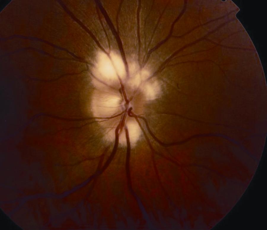
Glial veil.
Image credit: American Academy of Ophthalmology. Used with permission for educational use.

Glial veil.
Image credit: American Academy of Ophthalmology. Used with permission for educational use.

Glial tuft.
Image credit: American Academy of Ophthalmology. Used with permission for educational purposes.

Bergmeister papilla (histopathology). A, Gross photograph of infant eye showing fingerlike projection of whitish tissue (arrow) from the surface of the optic nerve head. B, Low-magnification photomicrograph of part A showing fibroglial tissue (arrow) projecting into the vitreous cavity from the optic nerve head. C, High-magnification photomicrograph of Bergmeister papilla demonstrating loose fibrous connective tissue with small capillary (arrow), surrounded by a thin layer of fibrous astrocyte-like cells (arrowhead).
Image credit: Courtesy of Robert H. Rosa, Jr., M.D. American Academy of Ophthalmology. Used with permission for educational purposes.

Bergmeister papilla.
Image credit: Courtesy of Kathleen Digre, M.D. Spencer S. Eccles Health Sciences Library, University of Utah, 2012. Neuro-ophthalmology Virtual Education Library. Available online. Used for educational purposes.


Crowded optic nerves. Note the anomalous vasculature and scalloped edges of the optic disc.
Image credit: Courtesy of William F. Hoyt, M.D. Spencer S. Eccles Health Sciences Library, University of Utah, 2002. Neuro-ophthalmology Virtual Education Library. Available online. Used for educational purposes.


Crowded optic nerve with glial remnant. Note the glial remnant in the right eye.
Image credit: Courtesy of William F. Hoyt, M.D. Spencer S. Eccles Health Sciences Library, University of Utah, 2002. Neuro-ophthalmology Virtual Education Library. Available online. Used for educational purposes.


Crowded congenital blurred disc. Note the anomalous vascular pattern and glial tissue on the disc in the right eye.
Image credit: Courtesy of William F. Hoyt, M.D. Spencer S. Eccles Health Sciences Library, University of Utah, 2002. Neuro-ophthalmology Virtual Education Library. Available online. Used for educational purposes.

Myelinated nerve fibers.
Image credit: Courtesy of Anthony C. Arnold, M.D. American Academy of Ophthalmology. Used with permission for educational purposes.

Myelinated nerve fibers.
Image credit: American Academy of Ophthalmology. Used with permission for educational purposes.

Myelinated nerve fibers
Image credit: American Academy of Ophthalmology. Used with permission for educational purposes.

Myelinated nerve fibers.
Image credit: Courtesy of Rod Foroozan, M.D. American Academy of Ophthalmology. Used with permission for educational purposes.

Myelinated nerve fibers.
Image credit: American Academy of Ophthalmology. Used with permission for educational purposes.

Myelinated nerve fibers.
Image credit: American Academy of Ophthalmology. Used with permission for educational purposes.

Myelinated nerve fibers.
Image credit: Courtesy of Andre Marques, M.D. American Academy of Ophthalmology. Used with permission for educational purposes.

Myelinated nerve fibers.
Image credit: American Academy of Ophthalmology. Used with permission for educational purposes.

Optic disc drusen.
Image credit: Courtesy of Andre Marques, M.D. American Academy of Ophthalmology. Used with permission for educational purposes.

Optic disc drusen.
Image credit: Courtesy of Rod Foroozan, M.D. American Academy of Ophthalmology. Used with permission for education purposes.

Optic disc drusen.
Image credit: Courtesy of Mansi Parikh, M.D. American Academy of Ophthalmology. Used with permission for educational purposes.

Buried optic disc drusen.
A. Left optic disc demonstrating fullness but no obvious drusen.
B. B-scan ultrasonography of patient’s left eye reveals hyperechoic, high signal characteristic of optic disc drusen (see arrow).
Image credit: American Academy of Ophthalmology. Used with permission for educational purposes.

Optic disc drusen.
Optic disc drusen are hyaline or calcified deposits within the optic nerve head. They may be visible at the optic nerve head or buried. Nasal visual field defects and other glaucoma-like visual field defects may be present, though most visual field defects are asymptomatic.
There are many imaging modalities that help visualize optic nerve drusen, including B-scan ultrasonography (C), autofluorescence (D), and CT (E).
A. Fundus photograph of optic disc drusen, showing blurred disc margin with scalloped edge, refractile bodies on the disc surface and at the superior pole, mild pallor, and no obscuration of retinal blood vessels.
B. Visual field patterns confirmed the presence of a nasal step produced by drusen involving the right disc.
C. B-scan ultrasonogram, demonstrating focal, highly reflective (due to calcification) elevation within the optic disc (arrow), which persists when the gain is decreased
D. Preinjection fundus photograph demonstrating autofluorescence (arrow).
E. CT scan of the orbits. Calcified optic disc drusen are visible bilaterally at the posterior globe–optic nerve junction (arrows).
Part A courtesy of Steven A. Newman, MD; part B courtesy of Michael S. Lee, MD; parts C, E courtesy of Anthony C. Arnold, MD; part D courtesy of Hal Shaw, MD.
Image credit: Basic and Clinical Science Course, Section 5: Neuro-Ophthalmology. San Francisco: American Academy of Ophthalmology, 2018-2019: 142. Used with permission for educational purposes.