Video credit: Lee AG. Pharmacologic Testing for Horner Syndrome. Video. YouTube. Available online. Accessed 02-27-2019.
Dilation Lag (video)
Video credit: Digre KB, Jacobson D, Balhorn R. Dilation Lag. Video. [Neuro-Ophthalmology Virtual Education Library: NOVEL Web Site]. 2005. Available online. Accessed 02-27-2019.
Horner Syndrome
Right Horner syndrome.
Note the anisocoria (right pupil smaller than left pupil) and right upper lid ptosis. There is not a prominent right lower lid ptosis present.
Image credit: American Academy of Ophthalmology. Used with permission for educational purposes.
Horner Syndrome
Right Horner syndrome.
Note the anisocoria (right pupil smaller than left pupil), and right upper eyelid ptosis. Right lower eyelid ptosis is not as pronounced in this case.
Image credit: American Academy of Ophthalmology. Used with permission for educational purposes.
Horner Syndrome
Right Horner syndrome.
Note the anisocoria (right pupil smaller than left pupil) and right upper and lower ptosis. In a child, the most common cause of congenital Horner syndrome is birth trauma to the brachial plexus, with acquired childhood Horner syndrome concerning for neuroblastoma. Apraclonidine should be avoided in small children due to the risk of CNS and respiratory depression.
Image credit: American Academy of Ophthalmology. Used with permission for educational purposes.
Horner Syndrome
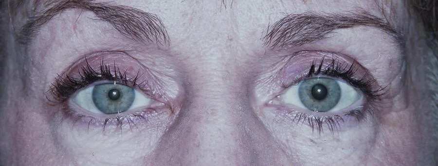
Left Horner syndrome.
Note the anisocoria (right pupil larger than left pupil) suggesting miosis of the left pupil. There is left upper lid ptosis and a very slight left lower lid ptosis, consistent with paralysis of the Müller and inferior tarsal muscles, respectively. There is also some mild increased conjunctival injection in the left eye, which can occur due to the disruption of sympathetic innervation to the periocular blood vessels.
Image credit: American Academy of Ophthalmology. Used with permission for educational purposes.
Hydroxyamphetamine Test
Hydroxyamphetamine test for Horner syndrome.
A. Before drops administered (suspected right Horner syndrome).
B. After drops administered. Note the dilation of both pupils. This indicates an intact 3rd-order, postganglionic neuron and localizes the lesion to the 1st-order (central) or 2nd-order (preganglionic) neuron.
Image credit: Modified from clinical images courtesy of Lanning B. Kline, M.D. American Academy of Ophthalmology. Used with permission for educational purposes.
Apraclonidine Test
Apraclonidine test for Horner syndrome.
A. Before drops administered (suspected left Horner syndrome).
B. After drops administered. Note the slight “reversal of anisocoria” in the left eye and the resolution of ptosis.
Image credit: Kanagalingam S, Miller NR. Eye Brain 2015;7:35-46. Available online. Used under the Creative Commons Attribution - Non Commercial (unported, v3.0) License.
Cocaine Test
Cocaine test for Horner syndrome.
A. Before drops administered (suspected right Horner syndrome).
B. After drops administered. Note that there is some pupil dilation in the right eye, but the amount of anisocoria is ≥1 mm.
Image credit: Kanagalingam S, Miller NR. Eye Brain 2015;7:35-46. Available online. Used under the Creative Commons Attribution - Non Commercial (unported, v3.0) License.


