hyperopia

Hyperopic Optic Nerve

Hyperopic optic nerve (Golnik, UpToDate).jpg

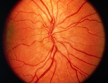
Hyperopic optic nerve. Note the crowded appearance of the optic nerve with nasal elevation. There is mild venous tortuosity, which can suggest venous outflow congestion and obscure the diagnosis. There is some anomalous branching of the retinal vessels, suggestive of pseudopapilledema. Despite the elevation and crowding of the optic disc, there is no whitening of the peripapillary retinal nerve fiber, no obscuration of the retinal vessels, no retinal or choroidal folds, and no nerve fiber layer hemorrhages.

Image credit: Karl Golnik, M.D. on UpToDate. Available online. Used for educational purposes.

Hyperopic Optic Nerve

Hyperopic optic nerve (Entokey).jpg

Hyperopic optic nerve. Note the generally ill-defined appearance to the optic nerve, with obscuration of the nasal contour. There is a small cup, and there may be a very slight elevation to the superior edge of the optic nerve. However, there is no whitening of the peripapillary nerve fiber layer, no obscuration of the retinal vessels, no venous engorgement or tortuosity, no true blurring of the nerve margins, no hemorrhages, and no retinal or choroidal folds.

Image credit: Appearance of optic nerve disorders. Entokey. Available online. Used for educational purposes.