Image credit: derm101.com.
There are a lot of practice questions on molluscum contagiosum. Although its clinical appearance and histopathology are fairly distinctive, I often confuse this with keratoacanthoma.
Gross Appearance
Image credit: drcharlesgeneslaw.files.wordpress.com.
- Molluscum contagiosum lesions are dome-shaped, waxy nodules with central umbilication.
Histopathology
Histopathology of molluscum contagiosum. Note the increased thickness of the surrounding epithelium (acanthosis). Within the epithelium there are eosinophilic inclusion bodies (Henderson-Patterson bodies), which become more and more basophilic as it rises to the surface.
Image credit: Wikipedia.
- The key histopathologic description for molluscum contagiosum is acanthosis with a central crater. This is nearly identical to the histopathologic description of a keratoacanthoma (acanthosis and hyperkeratosis with a central keratin-filled crater). One of the key differences is the amount of keratin - molluscum lesions do not have keratin in the crater (they have virus particles instead), keratoacanthomas do.
- Molluscum lesions have eosinophilic inclusion bodies, called Henderson-Patterson bodies. They become more and more basophilic as they migrate to the surface.
Key Facts
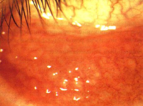
Molluscum contagiosum can cause follicular conjunctivitis, and should be considered in the differential diagnosis of follicular conjunctivitis.
Image credit: https://iliveok.com/sites/default/files/5_12.jpg
- Molluscum contagiosum is caused by a poxvirus.
- It can cause a follicular conjunctivitis.
- Multiple molluscum lesions should prompt investigation of an immunocompromised state, especially AIDS.
Management
- Molluscum lesions can be managed by observation, excision, curettage, or controlled cryotherapy.
References and Additional Reading
- Basic and Clinical Science Course, Section 4: Ophthalmic Pathology and Intraocular Tumors. American Academy of Ophthalmology, 2017-2018 edition.
- Basic and Clinical Science Course, Section 7: Orbit, Eyelids, and Lacrimal System. American Academy of Ophthalmology, 2017-2018 edition.
Do you have any suggestions on what else might be important to remember about molluscum contagiosum that may show up on the OKAP? Do you have any tips for helping to remember all of this information? Do you have any requests for specific topics to cover? Leave a comment or contact us!


