Anatomy

Binocular (Fusion) Eye Movements

Binocular (fusion) eye movements are synchronized eye movements that help maintain a clear and steady single image despite having two eyes, 12 extraocular motility muscles, and six cranial nerves controlling everything.

My residents often consider binocular eye movement-related problems and understanding the systems governing fusion as some of the more challenging problems encountered in ophthalmology and neuro-ophthalmology. The symptoms are often vague and difficult to describe. Furthermore, assessing and describing abnormal binocular eye movements are often subtle or challenging.

While ductions (ocular rotations) and alignment are often better conceptualized, abnormalities in binocular eye movements can be just as impactful to vision as strabismus or gaze palsy.

There are six different binocular eye movements:

  • Fixation

  • Saccades

  • Smooth pursuits

  • Vestibular-ocular reflex

  • Optokinetic system

  • Vergences

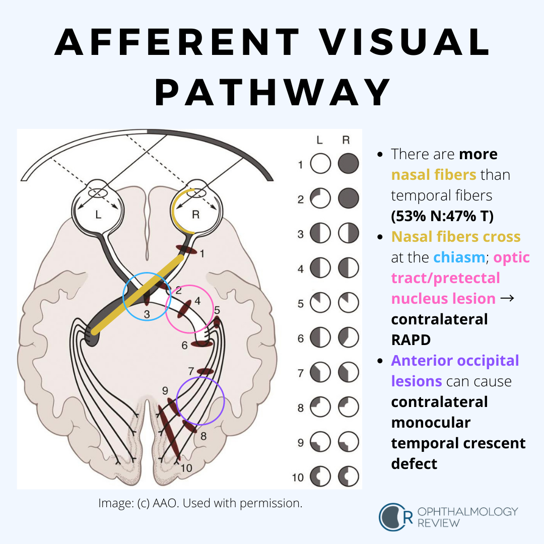
While the Basic and Clinical Science Course explains these systems in detail and shows the underlying pathways that govern each system (important for localization of lesions), I typically teach residents to consider these movements based on what the eyes are doing, what the head is doing, what the object of interest is doing, and how fast the movements are. All these movements are supranuclear (that is, the signals that control these movements are initiated before the cranial nerve nuclei are activated), so diseases that cause abnormalities in these eye movements affect cortical or brainstem structures rather than peripheral nerves or muscles.

Ciliary Ganglion

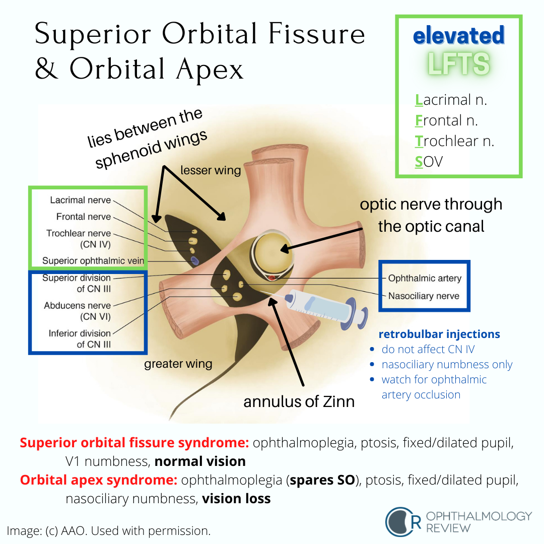
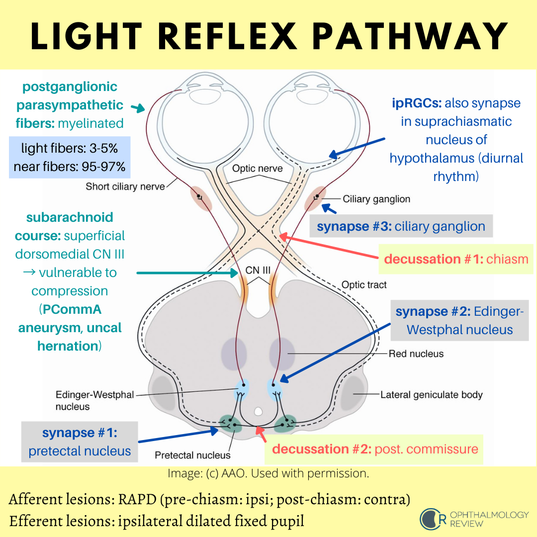
The ciliary ganglion serves as the site of synapse for the parasympathetic nerves innervating the eye.  Because of the many nerves that course through it (not all of them synapse!) and its anatomical location, this structure is of importance in learning the basics of ophthalmology.  According to the Basic and Clinical Science Course, it is located lateral to the ophthalmic artery, situated between optic nerve and lateral rectus muscle, approximately 1 cm (10 mm) anterior to the annulus of Zinn and 1.5-2 cm (15-20 mm) posterior to the globe (1-5).

Orbital And Eyelid Anatomy

I'm going to shift gears a little bit and start reviews on some of the other sections.  I originally had planned to go in order of the BCSC sections and follow the OKAP content outline, but I realized that of all the sections to cover, General Medicine is one of the smallest sections in terms of content to know.  So while I will likely get back to it sometime in the future, I wanted to make sure the key subjects were discussed prior to the test.

There are many facts in the Fundamentals and Principles of Ophthalmology section of the BCSC that will likely be tested as quick recall.  I promise, I will eventually provide numerous resources and tools to help remember these facts; for this article, I will try to cover the most important concepts.  I am intentionally leaving out details that may be more challenging to test (meaning I have a hard time coming up with a practice question about it).

Orbital Roof

The orbital roof separates the orbit from the anterior cranial fossa, which houses the frontal lobes of the brain.  There are several structures and features regarding the orbital roof that we need to remember.  While this article will try to list most of the important features of the orbital roof, it is by no means comprehensive.

Orbital Bones

There are 7 bones that comprise the orbit.  It is our job as ophthalmologists to be able to readily identify these bones and know pretty much every bump, notch, hole, and contour of these bones and what structures pass through, travel along, and attach to these bones.

Whitnall's Tubercle

The lateral orbital tubercle, or Whitnall's tubercle, is found on the zygomatic bone.  According to the Basic and Clinical Science Course, it is typically around 11 mm inferior to the frontozygomatic suture (the junction between the frontal bone and zygomatic bone) (1), and sits 4-5 mm posterior to the lateral orbital rim around the midline (2).